
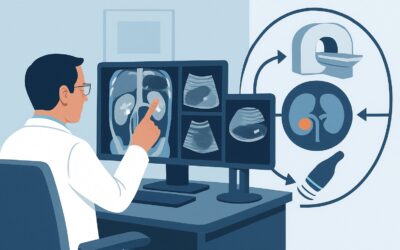
Which Imaging Is Best for Renal Cell Carcinoma Follow-up After Ablation?
ACR-guided imaging workflow for Follow-up for clinically localized renal cell carcinoma; post ablation. (Post Treatment Follow-up and Active Surveillance o
ACR-guided imaging workflow for Follow-up for clinically localized renal cell carcinoma; post ablation. (Post Treatment Follow-up and Active Surveillance o
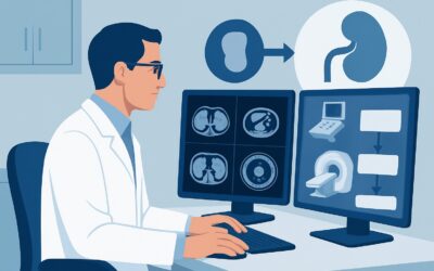
ACR-guided imaging workflow for Follow-up for clinically localized renal cell carcinoma; post radical or partial nephrectomy. (Post Treatment Follow-up and

ACR-guided imaging workflow for Palpable abdominal mass. Suspected abdominal wall mass. Initial imaging. (Palpable Abdominal Mass-Suspected Neoplasm): reco
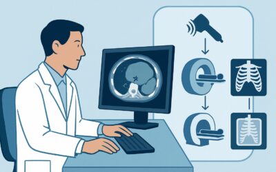
ACR-guided imaging workflow for Palpable abdominal mass. Suspected intra-abdominal neoplasm. Initial imaging. (Palpable Abdominal Mass-Suspected Neoplasm):
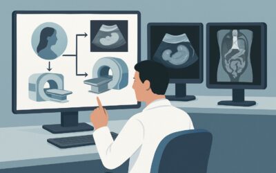
ACR-guided imaging workflow for Assessment of complications: GTD and GTN. (Gestational Trophoblastic Disease): recommended study, differential, downstream

ACR-guided imaging workflow for Surveillance of GTN, including refractory, relapsed, or quiescent GTN. (Gestational Trophoblastic Disease): recommended stu

ACR-guided imaging workflow for Staging and risk assessment: suspected or established diagnosis of gestational trophoblastic neoplasia (GTN). (Gestational
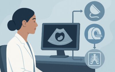
ACR-guided imaging workflow for Suspected or initial diagnosis of gestational trophoblastic disease (GTD). (Gestational Trophoblastic Disease): recommended
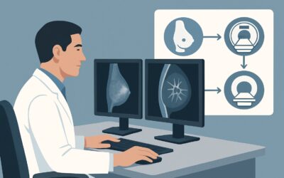
ACR-guided imaging workflow for Surveillance. Stage I breast cancer. Asymptomatic. Rule out bone, thoracic, abdominal, and brain metastases. Initial imagin

ACR-guided imaging workflow for Adult. Suspected or known hyperfunctioning pituitary adenoma (hyperthyroidism [high thyroid-stimulating hormone], Cushing s