

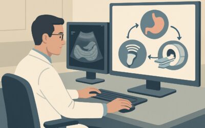
What Imaging Is Best for Epigastric Pain with Suspected Reflux, Gastritis, or Ulcer?
ACR-guided imaging workflow for Epigastric pain with clinical suspicion for acid reflux or esophagitis or gastritis or peptic ulcer or duodenal ulcer. Init
Part of the imaging decision system doctors were never taught to navigate at speed: which study to order, which protocol, and what the published appropriateness guidance actually says for GI imaging. Plain-English references, scenario by scenario.
ACR-guided imaging workflow for Epigastric pain with clinical suspicion for acid reflux or esophagitis or gastritis or peptic ulcer or duodenal ulcer. Init

ACR-guided imaging workflow for Suspected complication postproctectomy or coloproctectomy or colectomy with pouch or other anastomosis. Initial imaging. (A

ACR-guided imaging workflow for Suspected proctitis or pouchitis. Initial imaging. (Anorectal Disease): recommended study, differential, downstream pathway
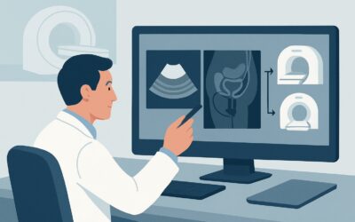
ACR-guided imaging workflow for Suspected rectal fistula. Rectovesicular or rectovaginal. Initial imaging. (Anorectal Disease): recommended study, differen
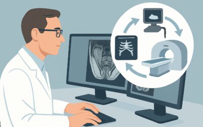
ACR-guided imaging workflow for Suspected perianal disease. Abscess or fistula. Initial imaging. (Anorectal Disease): recommended study, differential, down
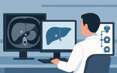
ACR-guided imaging workflow for Incidental liver lesion, less than 1 cm on US, noncontrast or single-phase CT, or noncontrast MRI. Known chronic liver dise

ACR-guided imaging workflow for Indeterminate, less than 1 cm liver lesion on initial imaging with CT (noncontrast or single-phase) or noncontrast MRI. Kno
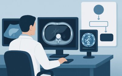
ACR-guided imaging workflow for Incidental liver lesion, greater than 1 cm on US, noncontrast or single-phase CT, or noncontrast MRI. Known chronic liver d
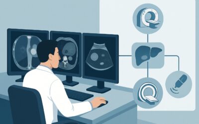
ACR-guided imaging workflow for Indeterminate, greater than 1 cm liver lesion on initial imaging with CT (noncontrast or single-phase) or noncontrast MRI.

ACR-guided imaging workflow for Indeterminate, greater than 1 cm liver lesion on initial imaging with CT (noncontrast or single-phase) or noncontrast MRI.