Should You Order a Radiograph or CT First for Stable Blunt Chest Trauma?
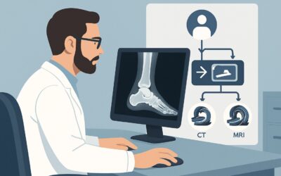
A 45-year-old restrained driver is brought to the emergency department after a high-speed motor vehicle collision. He is awake, alert, and his vitals are stable, but he complains of significant right-sided chest pain and has a prominent seatbelt sign across his upper torso. You hear decreased breath sounds on the right. You suspect a pneumothorax, rib fractures, or even a pulmonary contusion, but need to decide on the most appropriate initial imaging study to guide immediate management. This article details the clinical workflow for this specific scenario: an adult with major blunt trauma who is hemodynamically stable with suspected chest injury. According to the American College of Radiology (ACR) Appropriateness Criteria, a Radiography trauma series is rated as Usually Appropriate for this initial evaluation.
Who Fits This Clinical Scenario for Suspected Chest Trauma?
This guidance applies specifically to adult patients who have sustained major blunt force trauma and are hemodynamically stable, but where there is a clinical suspicion for thoracic injury. The key inclusion criteria are:
- Adult Patient: This workflow is not tailored for pediatric trauma, which has different considerations for radiation dose and injury patterns.
- Major Blunt Trauma: The mechanism of injury is significant, such as a motor vehicle collision, fall from height, or direct blow to the chest. This is not for minor bumps or bruises.
- Hemodynamically Stable: The patient has a normal blood pressure, heart rate, and is not in shock. This is a critical differentiator.
- Suspected Chest Trauma: Clinical signs point to the chest as a potential site of injury. This includes chest wall tenderness, crepitus, palpable fractures, a seatbelt sign, dyspnea, hypoxia, or abnormal lung sounds on auscultation.
It is crucial to distinguish this presentation from similar but distinct clinical situations that require a different approach. This guidance does not apply if:
- The patient is hemodynamically unstable. A hypotensive or tachycardic patient requires immediate resuscitation and often follows a different imaging algorithm, such as an eFAST exam followed by emergent intervention or a “pan-scan” CT. This falls under the Major Blunt Trauma variant for unstable patients.
- The trauma is isolated to another body part. If the mechanism and physical exam strongly suggest an isolated head, facial, or extremity injury without any signs of thoracic involvement, the imaging workup should be targeted to that specific area.
- The injury is penetrating. Gunshot wounds or stab wounds have entirely different injury patterns and imaging protocols.
What Diagnoses Are You Working Up in Blunt Chest Trauma?
When ordering initial imaging for a stable patient with suspected blunt chest trauma, you are evaluating for a range of injuries, from the common to the rare but life-threatening. The differential diagnosis drives the choice of imaging modality.
Pneumothorax and Hemothorax
These are among the most common and immediately consequential injuries. A pneumothorax (air in the pleural space) or hemothorax (blood in the pleural space) can compromise ventilation and circulation. A simple chest radiograph is often sufficient to detect a clinically significant collection that requires intervention, such as a chest tube.
Rib Fractures and Flail Chest
Fractures of the ribs are very common after blunt trauma. While a single, non-displaced fracture may be managed conservatively, multiple fractures can lead to significant pain, splinting, and atelectasis. A “flail chest,” where a segment of the chest wall moves paradoxically due to multiple fractures in multiple places, is a sign of severe trauma and can cause respiratory failure.
Pulmonary Contusion
This is a “bruise” of the lung tissue, leading to edema and hemorrhage within the alveoli. It can impair gas exchange and may not be fully apparent on the initial radiograph, often worsening over the first 24-48 hours. While CT is more sensitive, a radiograph can show early opacification suggestive of the diagnosis.
Aortic and Great Vessel Injury
Though less common, traumatic aortic injury is a highly lethal condition requiring emergent diagnosis and intervention. A chest radiograph is not sensitive enough to rule out this injury, but it can reveal suggestive secondary signs, such as a widened mediastinum, apical capping, or deviation of the trachea, which would mandate an immediate follow-up with a CTA (Computed Tomography Angiography) of the chest.
Why Is a Radiography Trauma Series Usually Appropriate for Initial Imaging?
For a hemodynamically stable adult with suspected blunt chest trauma, the ACR designates a Radiography trauma series as Usually Appropriate. This recommendation is based on a balance of diagnostic utility, speed, availability, and radiation exposure. The trauma series, typically including a portable anteroposterior (AP) chest view, provides a rapid and effective screen for the most immediate, life-threatening injuries.
A chest radiograph is highly effective for identifying large pneumothoraces, significant hemothoraces, multiple rib fractures, flail chest, and gross abnormalities of the mediastinal contour. Its primary advantage is speed and accessibility; a portable X-ray can be performed at the bedside in the trauma bay without moving a potentially unstable patient, providing critical information within minutes. The associated radiation dose is relatively low (ACR Relative Radiation Level ☢☢☢, 1-10 mSv).
While radiography is the recommended first step, other advanced imaging modalities are also rated for this scenario and play a crucial role in the downstream workflow:
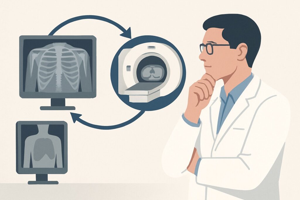
- CT chest with IV contrast and CTA chest with IV contrast are also rated Usually Appropriate. They are vastly more sensitive than radiography for detecting subtle pneumothoraces, pulmonary contusions, diaphragmatic injuries, and are the gold standard for evaluating for aortic or great vessel injury. However, they require transporting the patient to the CT scanner, involve a higher radiation dose (ACR RRL ☢☢☢, 1-10 mSv), and carry the risks of IV contrast administration. They are often used as the second step after an abnormal or inconclusive radiograph, or as the primary study in patients with a very high-risk mechanism of injury.
- US chest (ultrasound) is rated Usually not appropriate by the ACR as the definitive, comprehensive initial imaging study. While bedside ultrasound (as part of an eFAST exam) is invaluable for rapidly detecting pneumothorax and pericardial effusion, it is operator-dependent and does not provide a global assessment of the bony thorax, lung parenchyma, and mediastinum in the way that radiography or CT can.
The choice to start with radiography is a strategic one. It quickly triages patients, identifying those who need immediate procedures (like a chest tube) while selecting those who require the more detailed anatomical information provided by a subsequent CT scan.
What’s the Next Step After the Initial Chest Radiograph?
The results of the initial radiography trauma series will dictate the subsequent clinical and diagnostic workflow. The decision tree branches based on whether the findings are positive, negative, or indeterminate.
If the radiograph is clearly positive:
- Large Pneumothorax or Hemothorax: This finding prompts immediate intervention, typically with tube thoracostomy (chest tube insertion). Further imaging may be deferred until after the patient is stabilized.
- Widened Mediastinum or other signs of Aortic Injury: This is a radiological emergency. The patient should proceed directly to a CTA chest with IV contrast to definitively rule in or rule out an aortic or great vessel injury.
- Multiple Rib Fractures or Flail Chest: These patients require admission for pain management and respiratory monitoring. A follow-up CT chest with IV contrast is often warranted to fully assess for underlying injuries such as pulmonary contusion, laceration, or vascular damage that may not be visible on the plain film.
If the radiograph is negative but clinical suspicion remains high:
A normal chest radiograph does not exclude significant injury, particularly in the context of a high-energy mechanism (e.g., fall from height, high-speed collision) or persistent clinical signs (e.g., hypoxia, severe localized pain). In these cases, the next appropriate step is to obtain a CT chest with IV contrast. This provides a much more sensitive evaluation for occult pneumothorax, pulmonary contusion, and other subtle injuries.
If the radiograph is negative and clinical suspicion is low:
For patients with a lower-energy mechanism and improving symptoms, a negative chest radiograph may be sufficient. A period of observation followed by discharge with strict return precautions can be an appropriate course of action.
Common Pitfalls to Avoid in Stable Blunt Chest Trauma
Navigating the initial workup of blunt chest trauma requires vigilance to avoid common diagnostic errors. Be mindful of these potential pitfalls:
- Over-relying on a normal radiograph. A negative initial chest X-ray is reassuring but not definitive. Significant injuries like small pneumothoraces, pulmonary contusions, and especially aortic injuries can be missed. Always integrate the imaging findings with the mechanism of injury and the complete clinical picture.
- Forgetting the limitations of a supine film. In trauma, patients are often imaged in the supine position. This can obscure a pneumothorax (which collects anteriorly) and make it difficult to assess for pleural effusions. Look for subtle signs like the “deep sulcus sign” for a pneumothorax. If the patient’s spine is cleared and they are stable enough, an upright film is superior.
- Delaying definitive vascular imaging. If there is any radiographic or high-risk mechanistic concern for an aortic injury, do not delay obtaining a CTA of the chest. This is a time-sensitive, life-threatening diagnosis.
The most critical escalation point is any change in hemodynamic status. If a patient who was initially stable becomes hypotensive or tachycardic, you must immediately pivot from this workflow to an unstable trauma algorithm, which prioritizes resuscitation and rapid, focused assessment like the eFAST exam.
Related ACR Topics and Tools
This article focuses on one specific scenario. For a comprehensive overview of all clinical variants in this topic, or to explore the tools used to make these recommendations, please refer to the following resources.
- For breadth across all scenarios in Major Blunt Trauma, see our parent guide: Major Blunt Trauma: ACR Appropriateness Decoded.
- To explore other clinical situations, use the ACR Appropriateness Criteria Lookup tool.
- For details on imaging techniques, consult the Imaging Protocol Library.
- To discuss radiation exposure with patients, the Radiation Dose Calculator can help frame the conversation.
Frequently Asked Questions
Why is CT chest with contrast also rated ‘Usually Appropriate’ if radiography is the recommended first step?
Both are rated ‘Usually Appropriate’ because they are both valid initial choices depending on the specific clinical context. Radiography is often preferred first due to its speed, low cost, and bedside availability, making it an excellent triage tool. However, in a patient with a very high-risk mechanism of injury or multiple severe injuries, a provider might reasonably choose to proceed directly to CT as the initial study to get a more comprehensive and definitive assessment from the outset.
If my patient has a very high-risk mechanism, like a fall from >20 feet, should I skip the radiograph and go straight to CT?
Yes, this is often an appropriate clinical decision. While the ACR guidelines provide a framework, clinical judgment is paramount. For a hemodynamically stable patient with a severe mechanism of injury that raises high suspicion for aortic, spinal, or other internal injuries, proceeding directly to a CT of the chest (often as part of a whole-body or ‘pan-scan’ CT) is a common and justifiable pathway to avoid delays in diagnosing critical injuries.
Is there a role for a non-contrast chest CT in this scenario?
A non-contrast chest CT is rated ‘Usually Not Appropriate’ for the initial evaluation of major blunt trauma. Intravenous contrast is critical for assessing vascular structures (like the aorta), identifying active bleeding, and characterizing solid organ injuries. A non-contrast study would miss these key potential findings. The only common exception is if the patient has a severe contraindication to IV contrast, such as profound renal failure or a severe allergy, in which case the risks and benefits must be carefully weighed.
The eFAST exam was negative. Do I still need a chest radiograph?
Yes. The eFAST (Extended Focused Assessment with Sonography for Trauma) is excellent for rapidly identifying pneumothorax, hemothorax, and pericardial effusion at the bedside. However, it does not evaluate the bony thorax (ribs, sternum, spine), the lung parenchyma for contusion, or the mediastinum for signs of great vessel injury. A negative eFAST is reassuring for certain immediate life-threats, but a chest radiograph is still necessary for a more complete initial thoracic evaluation.
What specific findings on the initial radiograph should prompt an immediate CTA?
Any finding suggestive of a traumatic aortic or great vessel injury should trigger an immediate CTA of the chest. Key red flags include a widened mediastinum (typically >8 cm on a supine AP film), loss of the normal aortic knob contour, apical pleural capping, deviation of the trachea or nasogastric tube to the right, and depression of the left mainstem bronchus.
Reviewed by Pouyan Golshani, MD, Interventional Radiologist — May 29, 2026