

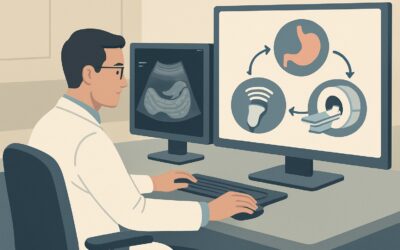
What Imaging Is Best for Epigastric Pain with Suspected Reflux, Gastritis, or Ulcer?
ACR-guided imaging workflow for Epigastric pain with clinical suspicion for acid reflux or esophagitis or gastritis or peptic ulcer or duodenal ulcer. Init
ACR-guided imaging workflow for Epigastric pain with clinical suspicion for acid reflux or esophagitis or gastritis or peptic ulcer or duodenal ulcer. Init

ACR-guided imaging workflow for Suspected complication postproctectomy or coloproctectomy or colectomy with pouch or other anastomosis. Initial imaging. (A

ACR-guided imaging workflow for Suspected proctitis or pouchitis. Initial imaging. (Anorectal Disease): recommended study, differential, downstream pathway
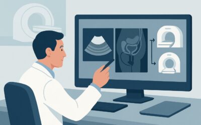
ACR-guided imaging workflow for Suspected rectal fistula. Rectovesicular or rectovaginal. Initial imaging. (Anorectal Disease): recommended study, differen
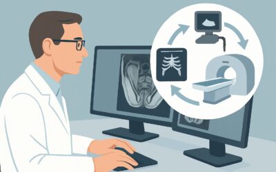
ACR-guided imaging workflow for Suspected perianal disease. Abscess or fistula. Initial imaging. (Anorectal Disease): recommended study, differential, down
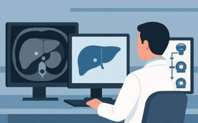
ACR-guided imaging workflow for Incidental liver lesion, less than 1 cm on US, noncontrast or single-phase CT, or noncontrast MRI. Known chronic liver dise

ACR-guided imaging workflow for Indeterminate, less than 1 cm liver lesion on initial imaging with CT (noncontrast or single-phase) or noncontrast MRI. Kno
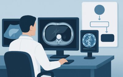
ACR-guided imaging workflow for Incidental liver lesion, greater than 1 cm on US, noncontrast or single-phase CT, or noncontrast MRI. Known chronic liver d
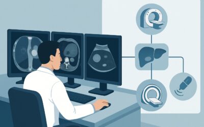
ACR-guided imaging workflow for Indeterminate, greater than 1 cm liver lesion on initial imaging with CT (noncontrast or single-phase) or noncontrast MRI.

ACR-guided imaging workflow for Indeterminate, greater than 1 cm liver lesion on initial imaging with CT (noncontrast or single-phase) or noncontrast MRI.