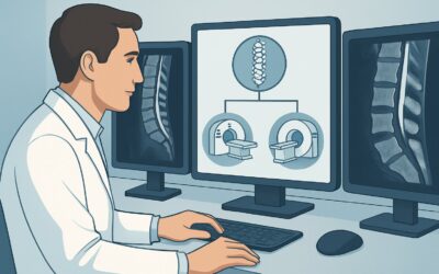
What Is the Best Initial Imaging for Acute Elbow or Forearm Pain in Adults?
A 48-year-old patient arrives at your clinic after a weekend of heavy yard work, complaining of new, sharp pain in his right elbow and proximal forearm. He doesn’t recall a specific fall but notes the pain worsened overnight. On examination, there is tenderness over the lateral epicondyle and radial head, with pain on resisted wrist extension, but no gross deformity or instability. You suspect a possible fracture or significant soft tissue injury, but the presentation is undifferentiated. The immediate clinical question is which imaging study to order first to guide management. This article provides a detailed workflow for this exact scenario, grounded in the American College of Radiology (ACR) guidelines. For the initial imaging of an adult with acute elbow or forearm pain, Radiography of the area of interest is rated Usually Appropriate.
Who Fits This Clinical Scenario for Acute Elbow Pain?
This guidance applies specifically to adult patients presenting with acute elbow or forearm pain (typically less than six weeks in duration) for whom initial, first-line imaging is being considered. The clinical picture is often undifferentiated, meaning a clear diagnosis of fracture versus soft tissue injury has not been established by history and physical exam alone. The patient may have a history of trauma (e.g., a fall, a direct blow) or the pain may be atraumatic in onset.
It is critical to distinguish this initial workup from subsequent imaging decisions. This workflow does not apply to patients who fall into these more specific categories:
- Suspected occult fracture with normal radiographs: If you have already obtained radiographs that are negative or indeterminate but maintain a high clinical suspicion for a fracture, you have moved to a different clinical scenario requiring a different imaging choice.
- Suspected soft tissue injury with normal radiographs: If initial radiographs have successfully ruled out a fracture and your primary concern is a tendon, ligament, or muscle injury, the next imaging step follows a separate decision pathway.
- Chronic elbow pain: Patients with pain lasting longer than six weeks are considered to have chronic pain, which involves a different differential diagnosis and imaging strategy.
This article is exclusively for the common, front-line decision: what is the single best first test for an adult with new elbow or forearm pain?
What Diagnoses Are You Working Up with Initial Elbow Radiographs?
Ordering initial radiographs is primarily a strategy to rapidly identify or exclude significant bony pathology. The differential diagnosis for acute elbow pain is broad, but the initial imaging is focused on the most urgent and structurally significant possibilities that would alter immediate management.
Fracture is the most common and consequential diagnosis to address. Radiographs are highly effective at identifying common fractures in this region, such as those involving the radial head or neck, the olecranon, or the distal humerus. A definitive diagnosis of a fracture immediately changes the treatment plan to immobilization, orthopedic consultation, or potential surgical intervention.
Joint Dislocation or Subluxation is another critical diagnosis that is readily apparent on radiographs. Assessing the alignment of the ulnohumeral and radiocapitellar joints is a primary goal of the initial study. An unrecognized dislocation can lead to severe long-term complications, including neurovascular injury and joint instability.
Occult Fracture Signs are a key finding in this scenario. In adults, the presence of a posterior fat pad sign or a displaced anterior fat pad (the “sail sign”) on a lateral radiograph is highly indicative of an intra-articular effusion. In the setting of trauma, this finding implies an occult, non-displaced intra-articular fracture (often of the radial head) even if a fracture line is not directly visible.
Underlying Bony Lesions or Arthritis may also be identified. While the pain is acute, radiographs can reveal pre-existing conditions like severe osteoarthritis, inflammatory arthritis, or (less commonly) a pathologic fracture through a bone lesion. These findings provide crucial context for the patient’s acute presentation.
Why Are Radiographs the Recommended First Study for Acute Elbow Pain?
The American College of Radiology designates Radiography of the area of interest as Usually Appropriate because it provides the highest diagnostic value for the lowest cost and risk in this initial setting. The rationale is built on efficiency, accessibility, and its power to effectively triage patients toward the correct management pathway.
Radiographs are fast, widely available, and inexpensive. They provide excellent spatial resolution for evaluating cortical bone, making them the ideal screening tool for fractures and dislocations. A standard two-view or three-view elbow series can quickly confirm a fracture, guiding immediate decisions on splinting and referral. Conversely, a negative radiograph that shows no effusion or bony abnormality significantly lowers the probability of a clinically significant fracture and allows the clinician to confidently manage the patient for a presumed soft tissue injury.
In contrast, more advanced imaging modalities are rated lower for this initial evaluation for clear reasons:
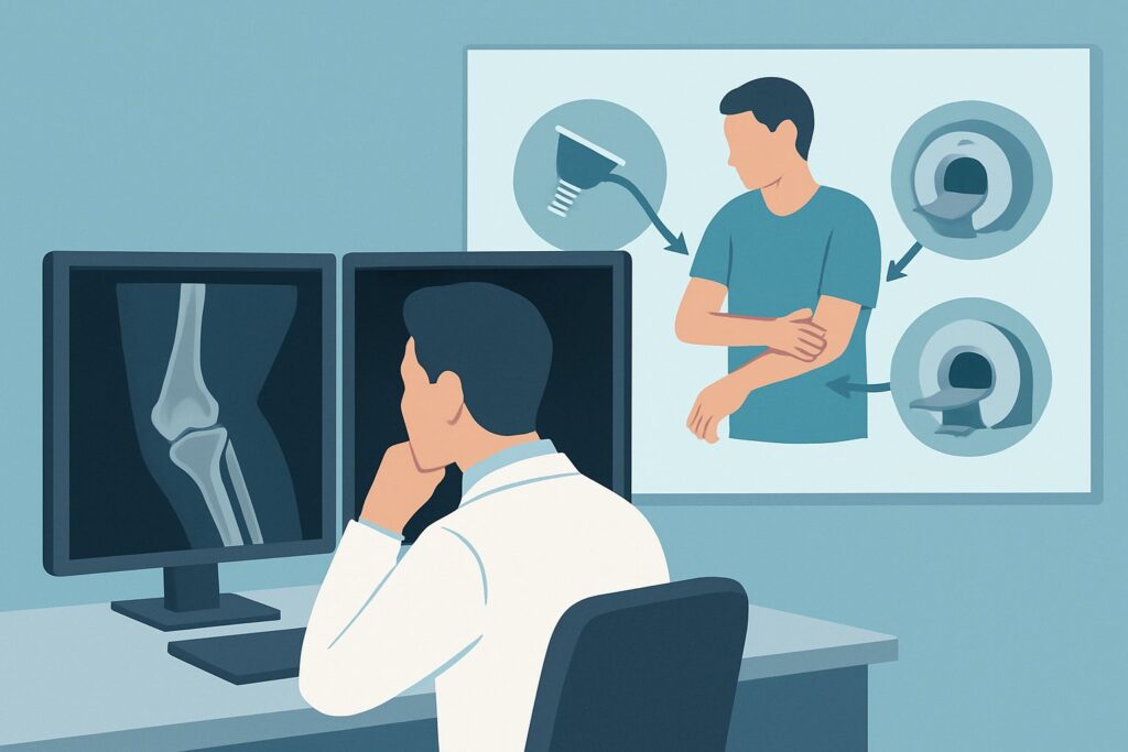
- MRI without IV contrast is rated Usually not appropriate. While MRI is the gold standard for evaluating soft tissues like ligaments and tendons, it is not the right first test for an undifferentiated presentation. It is more expensive, less available, and time-consuming. Ordering an MRI upfront to look for a fracture is inefficient; its role is as a problem-solving tool for suspected soft tissue injury after a fracture has been reasonably excluded by radiography.
- CT without IV contrast is also rated Usually not appropriate as a first-line test. CT offers superior detail for complex or occult fractures, but it comes with a higher radiation dose (RRL of Varies, but typically much higher than radiography) and cost. Its primary utility is in the next step of the workflow—characterizing a known complex fracture seen on a radiograph or evaluating for an occult fracture when radiographs are negative but clinical suspicion remains high.
The radiation dose for radiography is variable but generally low. No contrast is required. This combination of high diagnostic yield for the most urgent conditions (fracture, dislocation) and low risk makes it the definitive starting point for this clinical scenario.
What’s Next After Radiography? Downstream Workflow
The results of the initial radiograph create clear, divergent pathways for patient management. The next step is dictated entirely by the findings.
If the radiograph is positive for a fracture or dislocation: The diagnosis is made. The next step is orthopedic consultation. The complexity of the fracture seen on the radiograph will determine if further imaging, such as a CT scan, is needed for surgical planning, but this decision is typically made by the consulting specialist.
If the radiograph is negative for fracture but shows an effusion (e.g., a posterior fat pad sign): This is a high-suspicion finding for an occult, non-displaced intra-articular fracture (most commonly the radial head). The standard management is to treat the patient as if they have a fracture—typically with splinting and follow-up in 7-10 days. Repeat radiographs at follow-up may show a visible fracture line as bone resorption occurs. This finding places the patient into the sibling ACR scenario: “Adult. Acute elbow or forearm pain. Suspect fracture. Radiographs normal or indeterminate. Next imaging study.” In this context, CT or MRI may become appropriate if further clarification is needed.
If the radiograph is completely negative (no fracture, no effusion, normal alignment): A significant bony injury is highly unlikely. The patient can be managed for a soft tissue injury, such as a muscle strain, ligament sprain, or tendinopathy. This moves the patient into the other key sibling scenario: “Adult. Acute elbow or forearm pain. Suspect tendon or ligament or muscle injury. Radiographs normal or indeterminate.” If the patient fails to improve with conservative management, a follow-up evaluation with ultrasound or MRI may be warranted to assess the soft tissues.
Pitfalls to Avoid (and When to Get Help)
In the initial workup of acute elbow pain, several common pitfalls can delay diagnosis or lead to unnecessary imaging. Be mindful of the following:
- Under-reading the fat pads: Do not dismiss a visible posterior fat pad or a prominent “sail sign” on a lateral view. In an adult with trauma, this is a fracture until proven otherwise.
- Ordering MRI too early: Resist the urge to order an MRI as the initial test. It is an inefficient use of resources and often unnecessary if a radiograph can provide the diagnosis or effectively rule out a fracture.
- Inadequate radiographic views: Ensure a true lateral view is obtained. An oblique view can obscure a joint effusion and make fat pad assessment unreliable. If you are suspicious of a radial head fracture, dedicated radial head views can be helpful.
- Ignoring clinical context: If a patient has significant point tenderness directly over a bone (e.g., scaphoid, radial head) and the initial radiograph is negative, maintain a high index of suspicion for an occult fracture and manage the patient conservatively with immobilization and planned follow-up.
If there are any signs of neurovascular compromise, such as severe pain out of proportion, pallor, paresthesias, or pulselessness, this constitutes a clinical emergency requiring immediate orthopedic or surgical consultation, regardless of imaging findings.
Related ACR Topics and Tools
For a comprehensive overview of all clinical variants related to elbow and forearm pain, or to explore the tools used to develop this workflow, the following resources are available.
- For breadth across all scenarios in Acute Elbow and Forearm Pain, see our parent guide: Acute Elbow and Forearm Pain: ACR Appropriateness Decoded.
- To look up adjacent or alternative clinical scenarios, use the ACR Appropriateness Criteria Lookup tool.
- For detailed technical parameters of the recommended study, consult the Imaging Protocol Library.
- To discuss cumulative radiation exposure with patients, the Radiation Dose Calculator can help frame the conversation.
Frequently Asked Questions
Why not start with an ultrasound for acute elbow pain?
Ultrasound is rated ‘Usually not appropriate’ for the initial, undifferentiated evaluation of acute elbow pain. While it is excellent for evaluating specific soft tissue structures like tendons and ligaments, it is not a screening tool for bone. Radiography is far superior for identifying or excluding fractures and dislocations, which is the primary goal of the initial workup.
If a patient has a history of a fall, should I skip directly to CT?
No. Even with a clear history of trauma, radiography remains the appropriate first step. It is highly sensitive for the vast majority of fractures and has a much lower radiation dose than CT. CT is reserved for cases where the radiograph shows a complex fracture that needs better characterization for surgical planning, or when the radiograph is negative but clinical suspicion for an occult fracture remains very high.
What if the patient cannot fully extend their elbow for the radiograph?
Limited extension is common with elbow injuries due to pain and effusion. The technologist can often obtain two separate AP views—one with the forearm flat on the cassette and one with the humerus flat—to visualize the entire joint. A good quality lateral view is essential and can usually be obtained even with the elbow flexed at 90 degrees. This view is critical for assessing fat pads and joint alignment.
Does this guidance apply to children?
No, this specific workflow is for adults. While radiography is also the initial study of choice in children, the interpretation is more complex due to the presence of open growth plates (physes) and multiple ossification centers, which can be mistaken for fractures. Pediatric-specific guidelines and expertise are required.
Is there any role for a bone scan in this initial workup?
A bone scan is rated ‘Usually not appropriate’ for this scenario. While highly sensitive for detecting bony turnover and occult fractures, it is not specific and involves a significant radiation dose (1-10 mSv). It has been largely replaced by MRI or CT for evaluating suspected occult fractures due to their superior anatomic detail and, in the case of MRI, lack of ionizing radiation.
Reviewed by Pouyan Golshani, MD, Interventional Radiologist — May 30, 2026