What Is the Right Initial Imaging for a Stable Adult After Major Blunt Trauma?

A 34-year-old arrives in the emergency department after a high-speed motor vehicle collision. They are awake, alert, and their vital signs are stable, but the mechanism of injury is significant. You need to perform a thorough evaluation for occult injuries, but where do you start with imaging? The patient has no specific localizing complaints, just diffuse pain. This common scenario requires a systematic approach to initial imaging, balancing the need for rapid diagnosis against radiation exposure and resource utilization.
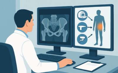
This article provides a detailed workflow for the initial imaging of a hemodynamically stable adult with major blunt trauma, not otherwise specified. For this specific presentation, the American College of Radiology (ACR) Appropriateness Criteria rate a Radiography trauma series as Usually Appropriate, serving as a critical first step in the diagnostic cascade.
Who Fits This Clinical Scenario for Major Blunt Trauma?
This guidance applies to a specific, well-defined patient population. Correctly identifying if your patient fits this scenario is the first step to ordering the right study.
Inclusion Criteria:
- Adult Patient: The patient is skeletally mature.
- Major Blunt Trauma: The mechanism of injury is significant enough to raise suspicion for multi-system injury. This includes events like high-speed motor vehicle collisions, falls from a significant height (typically >10 feet), or pedestrian-versus-vehicle incidents.
- Hemodynamically Stable: The patient shows no signs of shock. This is generally defined by a normal blood pressure (e.g., systolic >90 mm Hg), a heart rate that is not persistently tachycardic, and adequate peripheral perfusion.
- Not Otherwise Specified: The patient’s initial examination does not point to a single, isolated injury. There are no clear, overwhelming signs of severe head trauma, facial fractures, or a grossly deformed extremity that would immediately become the primary focus of the workup.
Exclusion Criteria (These Patients Follow a Different Workflow):
- Hemodynamically Unstable Patients: An unstable patient requires a different, more urgent algorithm, often beginning with a bedside FAST (Focused Assessment with Sonography for Trauma) exam to search for pericardial or intraperitoneal fluid. This is a distinct clinical scenario.
- Patients with Obvious, Localizing Injuries: If the patient has clear signs of isolated severe head trauma, facial injury, or extremity trauma, they would fit a more specific ACR variant. For example, a patient with suspected pelvic trauma or chest trauma would follow a dedicated imaging pathway.
What Diagnoses Are You Working Up in This Stable Polytrauma Patient?
In a stable patient with a major mechanism of injury, the goal of initial imaging is to rapidly screen for potentially life-threatening or limb-threatening injuries that may not be apparent on physical examination. The differential diagnosis is broad, covering critical injuries across multiple body cavities.
Cervical Spine Injury
Any significant blunt trauma carries a risk of cervical spine fracture or ligamentous injury, which can have devastating neurological consequences if missed. The initial physical exam can be unreliable in a patient with distracting injuries. Imaging serves as a crucial screening tool to clear the cervical spine or identify an injury that requires immobilization and further evaluation.
Thoracic Injury
The chest contains vital structures that are vulnerable to blunt force. A chest radiograph is essential for identifying a pneumothorax (which could become a tension pneumothorax), a hemothorax, significant rib fractures (which can be associated with underlying lung contusion), or a widened mediastinum, a potential sign of a catastrophic aortic injury.
Pelvic Fracture
The pelvis is a ring structure, and high-energy impacts can cause fractures that are not only painful but can also be a source of significant, occult hemorrhage. An unstable pelvic fracture is a surgical emergency. An initial anteroposterior (AP) radiograph of the pelvis is the standard screening test to assess the integrity of the pelvic ring.
Occult Abdominal or Retroperitoneal Injury
While the patient is hemodynamically stable, this stability can be tenuous. Injuries to solid organs like the spleen, liver, or kidneys can bleed slowly. The initial trauma series does not directly evaluate these organs, but it sets the stage for further, more advanced imaging if clinical suspicion remains high or if other injuries (like lower rib or pelvic fractures) are found.
Why Is a Radiography Trauma Series a Recommended First Step?
The ACR rates two different imaging strategies as Usually Appropriate for this scenario: a Radiography trauma series and a CT whole body with IV contrast. The choice between them often depends on the specific mechanism, clinical suspicion, and institutional protocols, but understanding the rationale for each is key.
A Radiography trauma series (ACR rating: Usually Appropriate) typically consists of an AP chest radiograph, an AP pelvis radiograph, and a lateral cervical spine view. This combination is a rapid, low-cost, and universally available method to screen for the most immediate threats. It can quickly identify a large pneumothorax, an unstable pelvic fracture, or a significant cervical spine malalignment. The associated radiation dose is moderate (ACR Relative Radiation Level ☢☢☢, 1-10 mSv).
A CT whole body with IV contrast (ACR rating: Usually Appropriate) is also a valid initial approach, often termed a “pan-scan.” It provides a far more detailed and comprehensive evaluation of the head, neck, chest, abdomen, and pelvis in a single acquisition. It is superior for detecting subtle solid organ injury, active vascular extravasation, and complex fracture patterns. However, this comes at the cost of a higher radiation dose (ACR Relative Radiation Level ☢☢☢☢, 10-30 mSv) and requires moving a potentially injured patient to the CT scanner and administering intravenous contrast.
The decision to start with radiographs versus proceeding directly to CT is a major branch point. Many trauma centers use criteria like the NEXUS criteria for C-spine clearance and clinical judgment to decide. Starting with radiographs can quickly rule out major injuries and potentially avoid a higher-dose CT scan in lower-risk patients.
Why Alternatives Are Rated Lower:
- **CT whole body without IV contrast** is rated Usually not appropriate. In the trauma setting, IV contrast is essential. It opacifies the blood vessels and enhances the solid organs, allowing for the detection of vascular injuries (e.g., dissection, pseudoaneurysm), active bleeding (extravasation), and parenchymal lacerations of the liver, spleen, and kidneys. A non-contrast scan would miss most of these critical findings.
- MRI of the abdomen and pelvis is also rated Usually not appropriate for initial imaging. MRI is time-consuming, sensitive to patient motion, and logistically challenging for a monitored polytrauma patient. While it is excellent for specific follow-up questions (e.g., ligamentous spine injury, detailed soft tissue evaluation), it is not a primary screening tool in the acute trauma bay.
What’s the Next Step After the Initial Trauma Radiographs?
The results of the initial radiography trauma series will guide the subsequent diagnostic workflow. This is not the end of the evaluation but rather the beginning of a branching decision tree.
- If the radiographs are positive: The findings dictate the next step.
- Abnormal Chest X-ray: A widened mediastinum necessitates an urgent CT angiography (CTA) of the chest to evaluate for aortic injury. A pneumothorax may require a chest tube. Multiple rib fractures may prompt a CT of the chest to assess for underlying lung contusion.
- Abnormal Pelvis X-ray: Any suspected pelvic fracture requires an orthopedic consultation and typically a CT of the abdomen and pelvis with IV contrast to better define the fracture pattern and rule out associated bladder, bowel, or vascular injuries.
- Abnormal C-spine X-ray: Any fracture or malalignment seen on the radiograph requires a definitive CT of the cervical spine.
- If the radiographs are negative: A negative trauma series is reassuring but does not definitively exclude all significant injuries, particularly intra-abdominal pathology. If the mechanism of injury was severe or if the patient has persistent abdominal tenderness, altered mental status, or other concerning signs, the next step is often a CT of the abdomen and pelvis with IV contrast. This is a common and appropriate escalation to ensure no occult solid organ or mesenteric injury is missed.
- If the findings are indeterminate: Occasionally, a radiograph may be equivocal (e.g., poor visualization of the C7-T1 junction on a C-spine film). In these cases, the next step is almost always CT for definitive evaluation.
Pitfalls to Avoid (and When to Get Help)
Navigating the initial workup of a major trauma patient requires vigilance to avoid common errors that can delay diagnosis or lead to missed injuries.
- Inadequate Radiographs: Insist on technically adequate images. A chest X-ray that doesn’t include the costophrenic angles or a C-spine film that doesn’t visualize the C7-T1 junction is incomplete and potentially misleading.
- Over-reassurance from a “Stable” Patient: Hemodynamic stability can be temporary. A patient with a splenic laceration may be stable for hours before decompensating. Maintain a high index of suspicion based on the mechanism of injury, not just the initial vital signs.
- Forgetting the Rest of the Body: The standard trauma series focuses on the chest, pelvis, and C-spine. Do not forget to perform a thorough secondary survey and consider dedicated imaging for extremities if there is pain, swelling, or deformity.
- Delaying Definitive Imaging: While radiographs are a good screening tool, do not delay proceeding to CT if clinical suspicion for intra-abdominal, thoracic, or complex spinal injury remains high.
If a patient who was initially stable begins to show signs of decompensation (falling blood pressure, rising heart rate), escalate immediately. This may involve initiating massive transfusion protocols and consulting trauma surgery and interventional radiology, as the patient may be transitioning to the “hemodynamically unstable” workflow.
Related ACR Topics and Tools
For a comprehensive overview of imaging in all major blunt trauma scenarios, from unstable patients to those with specific suspected injuries, please see our parent guide. For tools to help you apply these criteria in your practice, explore the resources below.
- For breadth across all scenarios in Major Blunt Trauma, see our parent guide: Major Blunt Trauma: ACR Appropriateness Decoded.
- ACR Appropriateness Criteria Lookup — for adjacent scenarios
- Imaging Protocol Library — for technique on the recommended study
- Radiation Dose Calculator — for cumulative dose conversations
Frequently Asked Questions
Why are both a radiography series and a whole-body CT scan rated ‘Usually Appropriate’?
The ACR recognizes that both are valid initial strategies for a stable polytrauma patient. A radiography trauma series is a rapid, lower-radiation screening tool to find immediate life threats. A whole-body CT is more sensitive and comprehensive but involves more radiation and logistics. The best choice often depends on the severity of the mechanism, institutional protocols, and clinical judgment after the primary survey.
If the initial trauma radiographs are negative, is the patient ‘cleared’?
No. A negative trauma series is reassuring for major thoracic, pelvic, and cervical spine bony injuries, but it does not rule out significant intra-abdominal injuries (like liver or spleen lacerations), ligamentous spine injuries, or subtle fractures. Further imaging with CT is often required if the mechanism of injury was significant or if the patient has persistent symptoms.
What is the role of the FAST exam in a hemodynamically stable patient?
While the FAST (Focused Assessment with Sonography for Trauma) exam is the cornerstone of evaluating an unstable trauma patient, its role in stable patients is more debated. A positive FAST in a stable patient would typically prompt an immediate CT scan. However, a negative FAST exam is not sensitive enough to rule out all significant injuries, and many centers proceed to CT based on mechanism and physical exam regardless of the FAST result in this population.
Should I order a CT of the whole body without IV contrast to save the patient’s kidneys?
No. For initial trauma evaluation, a CT without IV contrast is rated ‘Usually Not Appropriate.’ The risk of missing a life-threatening vascular or solid organ injury far outweighs the risk of contrast-induced nephropathy, which is very low in patients with normal baseline renal function who are adequately hydrated. Omitting contrast severely limits the diagnostic utility of the scan in this setting.
How does this guidance change if my stable patient has a specific complaint, like severe flank pain?
If a stable patient has a clear localizing sign or symptom, you may shift to a more focused ACR workflow. For example, significant flank pain and suspicion of renal injury would fall under the ‘Suspected urinary system trauma’ variant, which has its own specific imaging recommendations, often involving a CT with delayed excretory phase imaging.
Reviewed by Pouyan Golshani, MD, Interventional Radiologist — May 29, 2026