Which Imaging Study Best Measures Component Rotation in Painful Knee Arthroplasty?

A 62-year-old patient returns to your clinic six months after a total knee arthroplasty (TKA), complaining of persistent anterior knee pain and a feeling of instability, especially when climbing stairs. The surgical recovery was otherwise uneventful, and inflammatory markers are normal. Standard anteroposterior, lateral, and sunrise radiographs are obtained and show well-fixed components without fracture, subsidence, or significant polyethylene wear. You suspect the components may be malrotated, but the radiographs are insufficient to confirm this. The critical question is how to accurately and non-invasively measure the rotational alignment of the femoral and tibial components. For this specific clinical scenario, the American College of Radiology (ACR) Appropriateness Criteria rate a CT of the knee without IV contrast as Usually Appropriate.
Who Fits This Clinical Scenario for Measuring Knee Component Rotation?
This guidance applies to a specific subset of patients with a painful TKA. The key inclusion criteria are a patient with a history of total knee arthroplasty presenting with persistent pain, particularly patellofemoral in nature, where initial radiographs have already been performed and are non-diagnostic for common causes like loosening, fracture, or infection. The primary clinical suspicion driving the imaging choice is component malrotation.
It is crucial to distinguish this presentation from other causes of post-TKA pain, which follow different diagnostic pathways:
- If infection is the primary concern: Patients with systemic signs like fever, or local signs such as warmth, erythema, and effusion, coupled with elevated inflammatory markers (ESR/CRP), fit the suspected infection scenario, which has a distinct imaging workup.
- If aseptic loosening or osteolysis is suspected: If radiographs show progressive radiolucent lines or component migration, the workup is focused on confirming loosening or particle disease, a different clinical question.
- If there is a history of acute trauma: For patients with a recent fall or injury, the primary concern is a periprosthetic fracture, which also follows a separate ACR-guided workflow.
This article is exclusively for the patient whose symptoms and initial negative radiographs point toward an issue with component alignment as the likely source of pain.
What Diagnoses Are You Working Up When Measuring Component Rotation?
When ordering advanced imaging to assess component alignment after a TKA, the differential diagnosis is narrow and focused on the mechanical causes of patellofemoral dysfunction and instability.
The primary diagnosis under consideration is femoral component malrotation. This is a well-recognized cause of pain and dissatisfaction after TKA. When the femoral component is placed in excessive internal rotation relative to the native transepicondylar axis, it forces the patella to track laterally, leading to increased patellofemoral contact pressures, anterior knee pain, and potential instability or subluxation. This is the most common rotational problem leading to revision surgery.
A related but distinct diagnosis is tibial component malrotation. While less common, internal rotation of the tibial component relative to the tibial tubercle can also disrupt the knee’s extensor mechanism. This can alter the flexion-extension gap and contribute to pain and a sense of instability, sometimes in conjunction with femoral malrotation.
Finally, the investigation is working up patellofemoral maltracking and instability as the direct symptomatic consequence of the underlying component malrotation. The patient’s symptoms of pain, clicking, or giving way are the clinical expression of this abnormal mechanical interaction between the patella and the femoral trochlea. The imaging aims to quantify the root cause of this maltracking.
Why Is CT Knee without IV Contrast the Recommended Study for This Presentation?
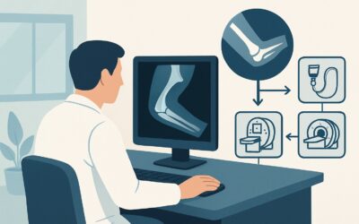
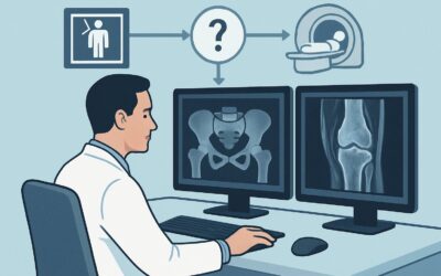
The ACR designates a CT of the knee without IV contrast as Usually Appropriate because it is the most accurate and reliable non-invasive method for quantifying the rotational alignment of TKA components.
The primary strength of CT is its ability to generate high-resolution, cross-sectional images of both the metallic implants and the surrounding bony anatomy. This allows for precise measurements using established protocols. Radiologists can accurately identify key anatomical landmarks that are obscured on plain radiographs, such as the surgical transepicondylar axis of the femur and the tibial tubercle. By comparing the orientation of the prosthetic components to these native landmarks, a precise measurement of rotational error can be calculated. Modern CT scanners employ sophisticated metal artifact reduction sequence (MARS) algorithms, which are critical for minimizing streak artifact from the implants and improving the visibility of the bone-implant interface and adjacent landmarks.
Alternative imaging modalities are rated lower for this specific clinical question:
- MRI knee without IV contrast is rated May be appropriate. While it offers excellent soft-tissue detail and avoids ionizing radiation (Radiation Level: O 0 mSv), it is significantly more susceptible to metal-induced artifacts than CT. These artifacts can severely distort the image around the prosthesis, making it difficult or impossible to identify the bony landmarks needed for accurate rotational measurements.
- Ultrasound is rated Usually not appropriate. Ultrasound waves cannot penetrate the metallic components of the arthroplasty, and the deep bony landmarks required for rotational assessment are not visible. It has no role in answering this specific clinical question.
From a safety and practical standpoint, the recommended CT is a low-dose examination (Relative Radiation Level: ☢ <0.1 mSv) and does not require intravenous contrast. This avoids the risks of contrast-induced nephropathy and allergic reactions, making it a safe and efficient choice.
What’s Next After the CT Scan? Downstream Workflow for Malrotation
The results of the CT scan directly guide the subsequent clinical management and create a clear decision-making pathway.
If the CT is positive for significant malrotation: When the CT report quantifies a degree of femoral or tibial component malrotation that is considered clinically significant and correlates with the patient’s symptoms, the next step is typically a consultation with an orthopedic surgeon specializing in revision arthroplasty. The objective measurements from the CT provide the surgeon with the data needed to plan a potential revision surgery to correct the alignment.
If the CT is negative for malrotation: If the scan demonstrates that both the femoral and tibial components are within the acceptable range of rotational alignment, malrotation is effectively ruled out as the cause of pain. The diagnostic focus must then shift to other potential etiologies. This may trigger a workup for a different clinical scenario, such as a suspected periprosthetic soft-tissue abnormality. The next logical imaging step could be an MRI without contrast (the May be appropriate study for this scenario) to evaluate for issues like tendinopathy, synovitis, or soft-tissue impingement. Non-surgical management, including physical therapy and targeted injections, would also be reconsidered.
If the results are borderline or indeterminate: In cases where the measured rotation is on the cusp of being clinically significant, the decision-making becomes more nuanced. The surgeon will need to carefully correlate the imaging findings with the patient’s specific symptoms and physical examination. A trial of conservative management, such as intensive physical therapy focused on quadriceps strengthening and patellar tracking, may be pursued before considering a revision.
Pitfalls to Avoid (and When to Get Help)
When ordering and interpreting imaging for TKA malrotation, several common pitfalls can compromise the diagnostic value of the study.
- Ordering a generic “Knee CT”: A standard knee CT protocol may not include the necessary axial slices through the femoral condyles (for the hip’s center of rotation) and the ankle mortise. The order must specify that the study is for TKA component rotation measurement to ensure the correct protocol is used.
- Ignoring metal artifact reduction: If the CT is performed on older equipment or without MARS algorithms, the resulting images may be non-diagnostic, wasting time and exposing the patient to unnecessary radiation. Confirm the imaging center uses an appropriate protocol.
- Over-relying on imaging alone: Rotational measurements exist on a spectrum. A finding of “borderline” malrotation requires careful clinical correlation. The decision for revision surgery should never be based solely on the CT report without a corroborating clinical picture.
If the clinical picture is complex or the imaging findings are equivocal, escalation to a subspecialist orthopedic surgeon with expertise in revision arthroplasty is the most appropriate next step.
Related ACR Topics and Tools
For a comprehensive overview of all clinical variants related to imaging after total knee arthroplasty, please consult our parent topic hub article. Additional tools are available to help select the right test and communicate with patients about radiation dose.
- For breadth across all scenarios in Imaging After Total Knee Arthroplasty, see our parent guide: Imaging After Total Knee Arthroplasty: ACR Appropriateness Decoded.
- To explore adjacent clinical scenarios and their recommended imaging pathways, use the ACR Appropriateness Criteria Lookup.
- For details on imaging techniques, refer to the Imaging Protocol Library.
- To discuss cumulative radiation exposure with your patients, the Radiation Dose Calculator can be a helpful resource.
Frequently Asked Questions
Why can’t standard X-rays be used to measure component rotation after a total knee arthroplasty?
Standard radiographs, including AP, lateral, and sunrise views, are two-dimensional projections and are not reliable for accurately measuring the three-dimensional rotational alignment of prosthetic components. While severe malrotation might be suspected, X-rays cannot provide the precise, reproducible measurements needed for surgical planning, which requires cross-sectional imaging like CT to compare component orientation to native bony landmarks.
Does the patient need IV contrast for a CT scan to evaluate TKA rotation?
No. Intravenous contrast is not necessary and is not recommended for this specific indication. The clinical question is about bony and component alignment, which is excellently visualized on a non-contrast CT. Omitting contrast avoids potential risks such as allergic reactions and contrast-induced nephropathy.
What specific information should I include in the order for this CT scan?
To ensure the correct protocol is performed, the order should explicitly state the indication. A good example would be: ‘CT knee without contrast for evaluation of painful total knee arthroplasty. Please perform TKA component rotational analysis protocol, including axial images through the hip and ankle for measurement.’
If the CT scan is negative for malrotation, what is the most likely cause of the patient’s pain?
If component malrotation is ruled out by CT, the differential for persistent anterior knee pain shifts to other causes. These can include soft-tissue impingement (e.g., patellar clunk syndrome), patellar tendinopathy, quadriceps dysfunction, complex regional pain syndrome, or referred pain. The next diagnostic step may involve an MRI to evaluate the soft tissues or a referral for further clinical evaluation by a specialist.
How much radiation is involved in a CT scan for TKA component rotation?
The recommended CT protocol is a low-dose examination. According to the ACR, the relative radiation level is minimal, rated as ☢ <0.1 mSv for an adult. This is a very low dose, comparable to or less than the radiation from several standard chest X-rays, and significantly less than the average annual background radiation in the United States.
Reviewed by Pouyan Golshani, MD, Interventional Radiologist — May 30, 2026