

Which Imaging Study Is Best for a Child with Suspected Orbital Infection?
ACR-guided imaging workflow for Child. Suspected orbital or periorbital infection. Initial imaging. (Orbital Imaging and Vision Loss-Child): recommended st
ACR-guided imaging workflow for Child. Suspected orbital or periorbital infection. Initial imaging. (Orbital Imaging and Vision Loss-Child): recommended st

ACR-guided imaging workflow for Child. Six months of age or older. Papilledema detected on the ophthalmologic examination or signs of raised intracranial p
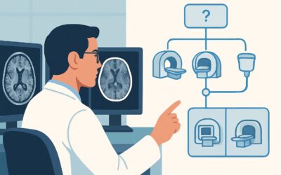
ACR-guided imaging workflow for Child. Vision loss and suspected optic pathway tumor, with or without a history of neurofibromatosis type 1. Initial imagin

ACR-guided imaging workflow for Child. Congenital or developmental abnormality leading to decreased visual acuity or vision loss. No leukocoria. Unilateral

ACR-guided imaging workflow for Child with isolated nystagmus. Initial imaging. (Orbital Imaging and Vision Loss-Child): recommended study, differential, d

ACR-guided imaging workflow for Child. Nontraumatic acute vision loss without papilledema. Initial imaging. (Orbital Imaging and Vision Loss-Child): recomm

ACR-guided imaging workflow for Child. Traumatic visual loss. Suspected orbital injury. Initial imaging. (Orbital Imaging and Vision Loss-Child): recommend

ACR-guided imaging workflow for Adult. Nonspecific dizziness without vertigo, ataxia, or other neurologic deficits. Initial imaging. (Dizziness and Ataxia)

ACR-guided imaging workflow for Adult. Chronic disequilibrium with signs of sensory or proprioceptive ataxia. Initial imaging. (Dizziness and Ataxia): reco

ACR-guided imaging workflow for Adult. Chronic disequilibrium with signs of cerebellar ataxia. Initial imaging. (Dizziness and Ataxia): recommended study,