
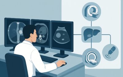
Which Imaging Best Characterizes a Liver Lesion in a Patient with Known Malignancy?
ACR-guided imaging workflow for Indeterminate, greater than 1 cm liver lesion on initial imaging with CT (noncontrast or single-phase) or noncontrast MRI.
ACR-guided imaging workflow for Indeterminate, greater than 1 cm liver lesion on initial imaging with CT (noncontrast or single-phase) or noncontrast MRI.

ACR-guided imaging workflow for Indeterminate, greater than 1 cm liver lesion on initial imaging with CT (noncontrast or single-phase) or noncontrast MRI.
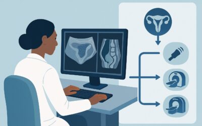
ACR-guided imaging workflow for Abnormal uterine bleeding. Follow-up imaging when surveillance is appropriate given findings from the initial ultrasound. (
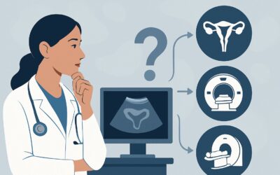
ACR-guided imaging workflow for Abnormal uterine bleeding. Follow-up imaging when original ultrasound is inconclusive or further imaging characterization i

ACR-guided imaging workflow for Abnormal uterine bleeding. Initial imaging. (Abnormal Uterine Bleeding): recommended study, differential, downstream pathwa
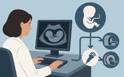
ACR-guided imaging workflow for Increased nuchal translucency in monochorionic twins at 11 to 14 weeks of gestation. (Nuchal Translucency Evaluation at 11
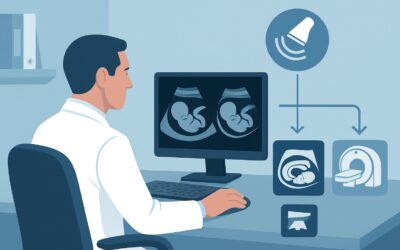
ACR-guided imaging workflow for Increased nuchal translucency in dichorionic twins at 11 to 14 weeks of gestation. (Nuchal Translucency Evaluation at 11 to

ACR-guided imaging workflow for Increased nuchal translucency measurement at 11 to 14 weeks of gestation for single gestation. (Nuchal Translucency Evaluat
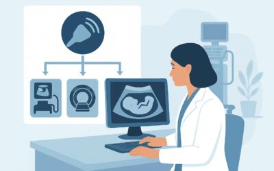
ACR-guided imaging workflow for Routine nuchal translucency measurement at 11 to 14 weeks of gestation for single or twin gestations. Initial imaging. (Nuc
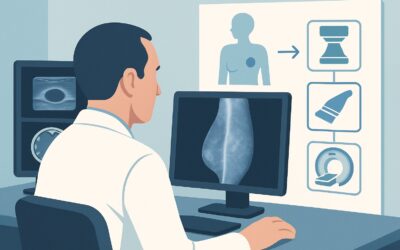
ACR-guided imaging workflow for Female. Palpable lump or clinically significant pain on the side of the mastectomy with reconstruction (autologous or nonau