
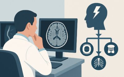
Which Imaging Study Is Best for Suspected Venous Injury After Head Trauma?
ACR-guided imaging workflow for Head trauma with suspected intracranial venous injury due to clinical risk factors or positive findings on prior imaging. (
Part of the imaging decision system doctors were never taught to navigate at speed: which study to order, which protocol, and what the published appropriateness guidance actually says for neuro imaging. Plain-English references, scenario by scenario.
ACR-guided imaging workflow for Head trauma with suspected intracranial venous injury due to clinical risk factors or positive findings on prior imaging. (

ACR-guided imaging workflow for Acquired conductive hearing loss secondary to cholesteatoma or neoplasm with suspected intracranial or inner ear extension.

ACR-guided imaging workflow for New-onset seizure. History of trauma. Initial imaging. (Seizures and Epilepsy): recommended study, differential, downstrea

ACR-guided imaging workflow for Anosmia or other abnormalities of the sense of smell (olfactory nerve, CN I). Initial imaging. (Cranial Neuropathy): recomm
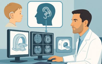
ACR-guided imaging workflow for Congenital hearing loss or total deafness or cochlear implant candidate. Surgical planning. (Hearing Loss and/or Vertigo):
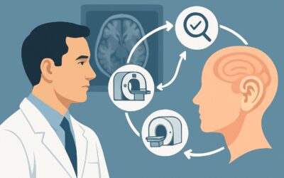
ACR-guided imaging workflow for Persistent vertigo with or without neurological symptoms (central vertigo). Initial imaging. (Hearing Loss and/or Vertigo):
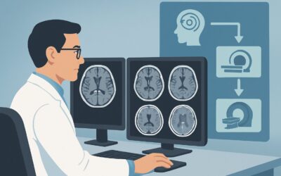
ACR-guided imaging workflow for Episodic vertigo with or without associated hearing loss or aural fullness (peripheral vertigo). Initial imaging. (Hearing
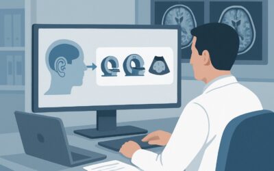
ACR-guided imaging workflow for Mixed conductive and sensorineural hearing loss. Initial imaging. (Hearing Loss and/or Vertigo): recommended study, differe
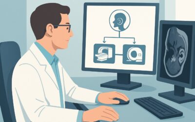
ACR-guided imaging workflow for Acquired sensorineural hearing loss. Initial imaging. (Hearing Loss and/or Vertigo): recommended study, differential, downs
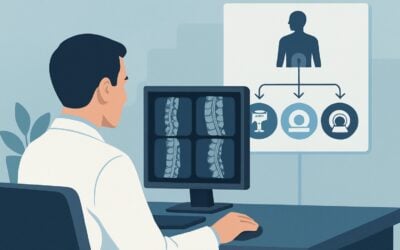
ACR-guided imaging workflow for Lumbosacral plexopathy, traumatic. Initial imaging. (Plexopathy): recommended study, differential, downstream pathway.