Should You Order MR Arthrography for Acute Shoulder Pain and a Suspected Labral Tear?
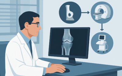
A 35-year-old patient presents to your clinic with acute, deep-seated right shoulder pain after a fall during a weekend softball game. They describe a painful “clicking” sensation with overhead movements. On examination, the O’Brien’s test is positive, raising suspicion for a superior labrum anterior-posterior (SLAP) tear. You’ve already obtained radiographs of the shoulder, which are negative for fracture or dislocation. Now, you face the critical decision: which advanced imaging study will definitively evaluate the glenoid labrum and guide the next steps in management? This article provides a focused workflow for this exact scenario, breaking down the rationale for the recommended imaging.
According to the American College of Radiology (ACR) Appropriateness Criteria, for an adult with acute shoulder pain, a physical examination consistent with a labral tear, and negative radiographs, MR arthrography shoulder is rated as Usually Appropriate.
Who Fits This Clinical Scenario for a Suspected Labral Tear?
This guidance is specifically for adult patients presenting with acute shoulder pain where the clinical suspicion for a glenoid labral tear is high. The key inclusion criteria are a compelling history (e.g., trauma, overhead throwing mechanism) and positive findings on specific physical examination maneuvers, such as the O’Brien’s test, crank test, or anterior apprehension test. A crucial prerequisite is that initial imaging with radiographs has already been performed and is either negative or indeterminate, effectively ruling out an obvious fracture or dislocation.
This workflow does not apply to several similar presentations. If the primary clinical suspicion is for a rotator cuff tear based on weakness with external rotation or a positive drop arm test, the imaging pathway may differ. Similarly, if radiographs are positive for a proximal humerus or clavicle fracture, the workup shifts to characterizing the fracture for surgical planning. This guidance is also distinct from the workup for chronic shoulder instability or when an occult fracture is the primary concern after trauma. Correctly identifying your patient’s presentation is the first step to ordering the most effective imaging study.
What Diagnoses Are You Working Up in This Scenario?
When ordering advanced imaging after negative radiographs in a patient with suspected labral pathology, you are investigating a specific set of intra-articular injuries that standard x-rays cannot visualize. The differential diagnosis is focused and drives the choice of a high-resolution, joint-distending imaging technique.
The primary diagnosis under consideration is a glenoid labral tear. This fibrocartilaginous ring is crucial for shoulder stability, and tears can occur from acute trauma or repetitive microtrauma. Common patterns include SLAP tears at the superior labrum or Bankart lesions associated with anterior instability. The physical exam findings are suggestive, but imaging is required for confirmation, characterization, and to guide potential surgical intervention.
Closely associated with labral tears are injuries to the glenohumeral ligaments. These ligaments are thickenings of the joint capsule and are critical static stabilizers. An MR arthrogram is highly effective at delineating these structures and identifying sprains or complete tears that often coexist with labral pathology and contribute to instability.
Another important consideration is an articular cartilage injury or chondral defect. Damage to the smooth cartilage lining the glenoid or humeral head can be a significant source of pain and mechanical symptoms. These defects are often subtle and best visualized when outlined by intra-articular contrast.
Why Is MR Arthrography the Recommended Study for a Suspected Labral Tear?
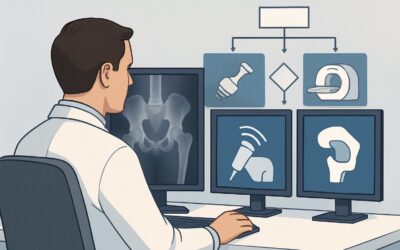
For this specific clinical question, the ACR panel rates three studies as Usually Appropriate: MR arthrography, MRI without contrast, and CT arthrography. However, MR arthrography is often considered the most definitive non-operative study for evaluating the labrum and associated stabilizing structures.
The core advantage of MR arthrography shoulder is the injection of dilute gadolinium-based contrast directly into the glenohumeral joint. This technique distends the joint capsule, separating intra-articular structures and forcing contrast into subtle tears of the labrum or rotator cuff that might otherwise be invisible. This provides superior delineation of the labrum’s morphology, attachment, and integrity, making it highly sensitive and specific for detecting and characterizing tears. Furthermore, it involves no ionizing radiation (0 mSv).
MRI shoulder without IV contrast is also rated Usually Appropriate. A non-arthrographic MRI can be sufficient, particularly on a high-field strength (3T) magnet, and avoids the invasive joint injection. However, its sensitivity for detecting non-displaced labral tears or evaluating the glenohumeral ligaments can be lower than that of an arthrogram, as a native joint effusion may not be present to outline the pathology.
CT arthrography shoulder is the third Usually Appropriate option. It provides excellent detail of the labrum and osseous structures. However, it delivers a significant radiation dose (ACR RRL® ☢☢☢☢, 10-30 mSv) and offers less soft tissue contrast for evaluating surrounding muscles and tendons compared to MRI. It is primarily reserved for patients with contraindications to MRI, such as incompatible implants or severe claustrophobia.
Studies like US shoulder are rated Usually not appropriate for this indication. While ultrasound is excellent for evaluating the rotator cuff tendons, it cannot adequately visualize deep intra-articular structures like the glenoid labrum. Once you’ve decided on the most appropriate MRI study, our protocol guide covers the essential techniques and reading principles. For more details, see our guide: MRI Shoulder Without Contrast.
What’s Next After MR Arthrography? Downstream Workflow
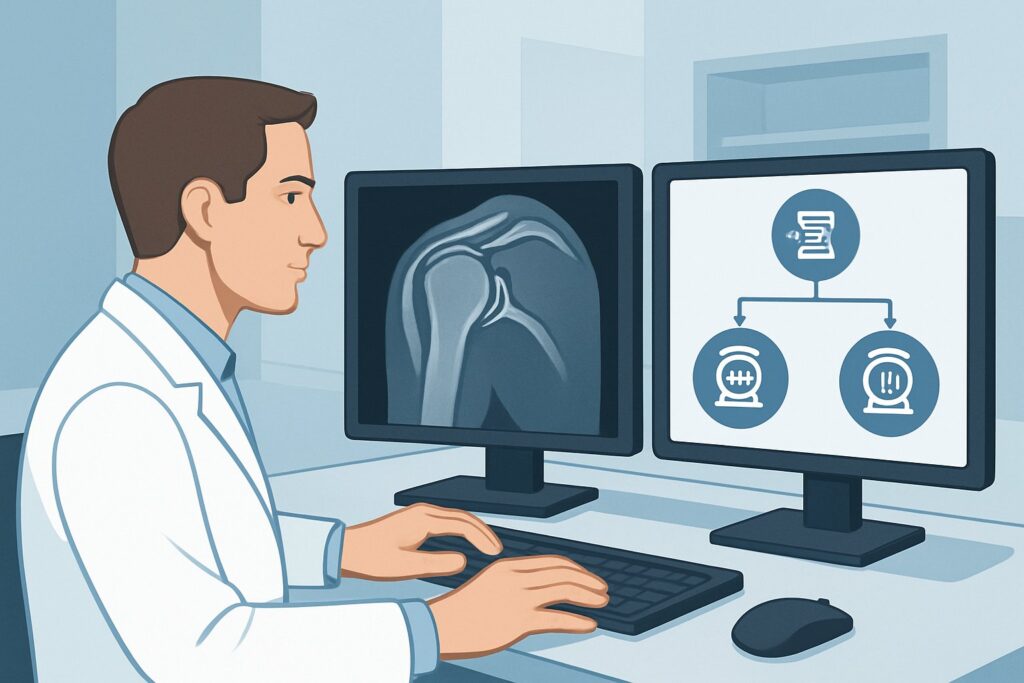
The results of the MR arthrogram will directly guide the subsequent clinical pathway, branching into non-operative and operative management strategies. Understanding these downstream decisions is key to effective patient counseling and care coordination.
If the study is positive for a significant labral tear (e.g., a displaced SLAP tear or a Bankart lesion with associated capsular injury) in a young, active patient, the next step is typically a referral to an orthopedic surgeon. The imaging findings will be critical for pre-operative planning, helping the surgeon anticipate the extent of the repair needed. The patient’s functional demands, symptoms, and goals will determine whether surgical stabilization is pursued.
If the MR arthrogram is negative and rules out a significant labral, ligamentous, or chondral injury, the focus shifts to non-operative management. This typically involves a structured physical therapy program aimed at strengthening the dynamic stabilizers of the shoulder (the rotator cuff and periscapular muscles). Anti-inflammatory medications and activity modification are also mainstays of treatment. A negative high-quality study provides confidence in pursuing a conservative course.
If the findings are indeterminate or show pathology that doesn’t fully explain the patient’s symptoms, a re-evaluation is warranted. This may involve considering other pain generators, such as biceps tendon pathology or nerve-related issues, and potentially consulting with a musculoskeletal radiologist to review the images. In some cases, a diagnostic injection or a second opinion from an orthopedic specialist may be necessary to clarify the diagnosis.
Pitfalls to Avoid (and When to Get Help)
Navigating the workup for a suspected labral tear requires careful attention to detail to avoid common missteps. A primary pitfall is ordering a non-arthrographic MRI when the pre-test probability for a labral tear is very high; this can lead to a false-negative result, delaying definitive diagnosis and treatment. Another error is relying on ultrasound for this specific indication, as it lacks the sensitivity for deep intra-articular structures. Also, ensure the patient has no contraindications to MRI or gadolinium contrast before ordering the study. If the clinical picture suggests glenohumeral instability but the MR arthrogram is equivocal, escalation to an orthopedic surgeon for their clinical correlation and potential examination under anesthesia may be the appropriate next step.
Related ACR Topics and Tools
This article focuses on a single, specific clinical scenario. For a comprehensive overview of all common presentations of acute shoulder pain and their corresponding imaging recommendations, please refer to our parent guide. To explore other scenarios or refine your understanding of imaging protocols and radiation safety, the following resources are available.
- For breadth across all scenarios in Acute Shoulder Pain, see our parent guide: Acute Shoulder Pain: ACR Appropriateness Decoded.
- ACR Appropriateness Criteria Lookup — for adjacent scenarios
- Imaging Protocol Library — for technique on the recommended study
- Radiation Dose Calculator — for cumulative dose conversations
Frequently Asked Questions
Why is MR arthrography preferred over a standard MRI for a suspected labral tear?
MR arthrography involves injecting contrast directly into the joint, which distends the capsule and outlines the labrum. This process forces contrast into small or non-displaced tears, significantly increasing the sensitivity for detection compared to a standard (non-arthrographic) MRI, where a joint effusion may not be present to provide natural contrast.
Is a 3T MRI without contrast good enough to rule out a labral tear?
A high-field strength 3-Tesla (3T) MRI without contrast is also rated ‘Usually Appropriate’ by the ACR and can have excellent diagnostic accuracy for labral tears, approaching that of arthrography in some studies. However, for the most subtle tears or in cases where clinical suspicion is very high, MR arthrography remains the gold standard for its superior ability to delineate intra-articular anatomy.
When should I choose CT arthrography instead of MR arthrography?
CT arthrography is an excellent alternative and is also rated ‘Usually Appropriate.’ It should be considered for patients who have contraindications to MRI, such as an incompatible pacemaker, certain cochlear implants, or severe claustrophobia. While it provides superb osseous and labral detail, it involves significant ionizing radiation and offers less soft tissue detail than MRI.
What if the patient’s pain is more consistent with a rotator cuff tear than a labral tear?
If the physical examination points primarily to a rotator cuff tear (e.g., weakness with external rotation, positive drop arm or empty can tests), the clinical scenario is different. For that presentation, both MRI without contrast and ultrasound are rated ‘Usually Appropriate’ as the initial advanced imaging studies. Ultrasound is particularly effective and cost-efficient for evaluating rotator cuff tendons.
Do I need to order any specific sequences for the MR arthrogram?
You do not typically need to specify individual sequences when ordering. The ordering physician should clearly state the clinical indication: ‘Acute shoulder pain, rule out labral tear.’ This allows the radiology department to perform their standardized shoulder arthrogram protocol, which will include the necessary fat-suppressed T1-weighted sequences in multiple planes and often an ABER (Abduction and External Rotation) view to best assess the anteroinferior labrum.
Reviewed by Pouyan Golshani, MD, Interventional Radiologist — May 30, 2026