
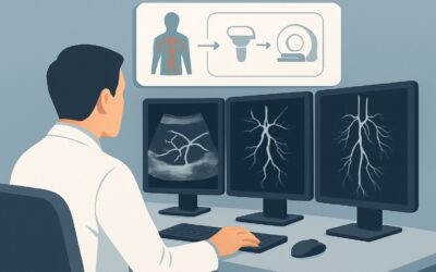
Which Imaging Study Is Best for Suspected Medium-Vessel Vasculitis (MVV)?
ACR-guided imaging workflow for Suspected medium-vessel vasculitis (MVV). Initial imaging. (Noncerebral Vasculitis): recommended study, differential, downs
ACR-guided imaging workflow for Suspected medium-vessel vasculitis (MVV). Initial imaging. (Noncerebral Vasculitis): recommended study, differential, downs
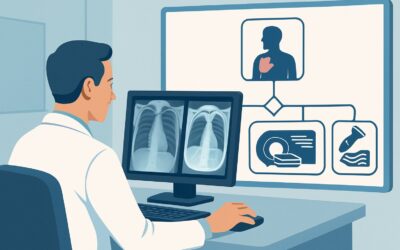
ACR-guided imaging workflow for Nontraumatic chest wall pain. History of prior chest intervention. Secondary evaluation after normal chest radiograph. Nex

ACR-guided imaging workflow for Nontraumatic chest wall pain. Suspected infectious or inflammatory condition. Secondary evaluation after normal chest radio

ACR-guided imaging workflow for Nontraumatic chest wall pain. Known or suspected malignancy. Secondary evaluation after normal chest radiograph. Next imagi
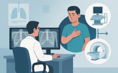
ACR-guided imaging workflow for Nontraumatic chest wall pain. No history of malignancy. Initial imaging. (Nontraumatic Chest Wall Pain): recommended study,

ACR-guided imaging workflow for Confirmed diffuse lung disease without acute clinical deterioration. Routine follow-up imaging clinically indicated. (Diffu

ACR-guided imaging workflow for Confirmed diffuse lung disease. Suspected acute exacerbation or acute deterioration. Initial Imaging. (Diffuse Lung Disease

ACR-guided imaging workflow for Suspected diffuse lung disease. Initial Imaging. (Diffuse Lung Disease): recommended study, differential, downstream pathwa

ACR-guided imaging workflow for Chronic cough lasting more than 8 weeks. Persistent symptoms despite initial clinical evaluation and empiric treatment. Ini
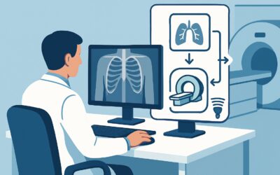
ACR-guided imaging workflow for Chronic cough lasting more than 8 weeks. Increased risk for lung cancer. Initial imaging. (Chronic Cough): recommended stud