

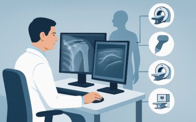
What Imaging Is Best for Suspected Biceps Tendon Pain After Normal X-Rays?
ACR-guided imaging workflow for Chronic shoulder pain. Suspect biceps tendon abnormality. Initial radiographs normal or inconclusive. Next imaging study. (
Part of the imaging decision system doctors were never taught to navigate at speed: which study to order, which protocol, and what the published appropriateness guidance actually says for MSK imaging. Plain-English references, scenario by scenario.
ACR-guided imaging workflow for Chronic shoulder pain. Suspect biceps tendon abnormality. Initial radiographs normal or inconclusive. Next imaging study. (

ACR-guided imaging workflow for Chronic shoulder pain. Suspect adhesive capsulitis. Initial radiographs normal or inconclusive. Next imaging study. (Chroni

ACR-guided imaging workflow for Chronic shoulder pain. Suspect labral pathology or shoulder instability. Initial radiographs normal or inconclusive. Next i
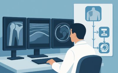
ACR-guided imaging workflow for Chronic shoulder pain. Initial imaging. (Chronic Shoulder Pain): recommended study, differential, downstream pathway.

ACR-guided imaging workflow for Soft tissue mass. Nondiagnostic radiograph and noncontrast enhanced ultrasound. MRI contraindicated. Next imaging study. (S
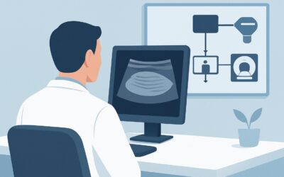
ACR-guided imaging workflow for Superficial soft tissue mass. Initial imaging. (Soft Tissue Masses): recommended study, differential, downstream pathway.
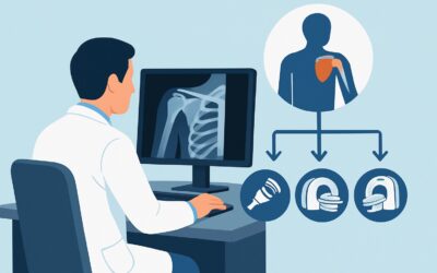
ACR-guided imaging workflow for Adult. Acute shoulder pain. Any etiology. Initial imaging. (Acute Shoulder Pain): recommended study, differential, downstre

ACR-guided imaging workflow for Chronic hip pain. Suspect noninfectious extra-articular abnormality, such as tendonitis or bursitis. Radiographs negative o

ACR-guided imaging workflow for Adult. Suspect stress fracture, excluding vertebrae. Initial imaging. (Stress (Fatigue/Insufficiency) Fracture, Including S

ACR-guided imaging workflow for Premenopausal females with risk factors. Males less than 50 years of age with risk factors. Follow-up to low bone mineral d