


How Should You Image Patients with Osteopenia and Added Vertebral Fracture Risk?
ACR-guided imaging workflow for Follow-up imaging. Patients with T-scores less than −1.0 (by DXA) and one or more of the following: 1) Females equal to or
Part of the imaging decision system doctors were never taught to navigate at speed: which study to order, which protocol, and what the published appropriateness guidance actually says for MSK imaging. Plain-English references, scenario by scenario.
ACR-guided imaging workflow for Follow-up imaging. Patients with T-scores less than −1.0 (by DXA) and one or more of the following: 1) Females equal to or
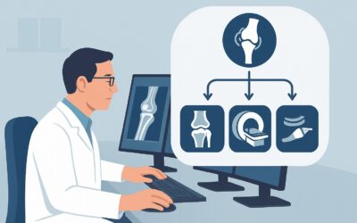
ACR-guided imaging workflow for Suspected osteomyelitis or septic arthritis or soft tissue infection (excluding spine and diabetic foot). Initial imaging.
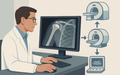
ACR-guided imaging workflow for Symptomatic patient with a primary shoulder arthroplasty, infection not excluded. Additional imaging following radiographs.
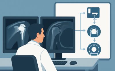
ACR-guided imaging workflow for Symptomatic patient with a primary shoulder arthroplasty, infection excluded. Suspected loosening. Additional imaging follo

ACR-guided imaging workflow for Symptomatic patient with a primary shoulder arthroplasty. Initial imaging. (Imaging After Shoulder Arthroplasty): recommend

ACR-guided imaging workflow for Routine follow-up of the asymptomatic patient with a primary shoulder arthroplasty. (Imaging After Shoulder Arthroplasty):

ACR-guided imaging workflow for Known axial spondyloarthritis. Follow-up for treatment response or disease progression. (Inflammatory Back Pain: Known or S

ACR-guided imaging workflow for Axial spondyloarthritis with spine ankylosis. Suspected fracture. Initial imaging. (Inflammatory Back Pain: Known or Suspec

ACR-guided imaging workflow for Inflammatory back pain. Suspected axial spondyloarthritis. Negative radiographs and negative MRI of the sacroiliac joints.

ACR-guided imaging workflow for Inflammatory back pain. Suspected axial spondyloarthritis. Initial imaging. (Inflammatory Back Pain: Known or Suspected Axi