

How Should You Assess Osteoporosis in Patients with Severe Spinal Degeneration?
ACR-guided imaging workflow for Males and females greater than or equal to 50 years of age. Suspected osteoporosis. Advanced degenerative changes of the sp
ACR-guided imaging workflow for Males and females greater than or equal to 50 years of age. Suspected osteoporosis. Advanced degenerative changes of the sp

ACR-guided imaging workflow for Initial imaging for premenopausal females or males less than 50 years of age. Individual with risk factors that could alter
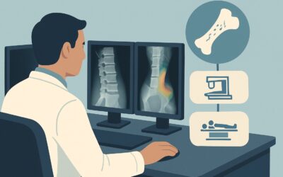
ACR-guided imaging workflow for Osteoporosis screening or initial imaging of clinically suspected low bone mineral density. (Osteoporosis and Bone Mineral

ACR-guided imaging workflow for Child. Chronic nonprogressive ataxia. Initial imaging. (Ataxia-Child): recommended study, differential, downstream pathway.

ACR-guided imaging workflow for Child. Chronic progressive ataxia. Initial imaging. (Ataxia-Child): recommended study, differential, downstream pathway.

ACR-guided imaging workflow for Child. Recurrent ataxia with interval normal neurology examination. Initial imaging. (Ataxia-Child): recommended study, dif

ACR-guided imaging workflow for Child. Acute ataxia, history of recent trauma. Initial imaging. (Ataxia-Child): recommended study, differential, downstream

ACR-guided imaging workflow for Child. Acute ataxia, no history of recent trauma. Initial imaging. (Ataxia-Child): recommended study, differential, downstr

ACR-guided imaging workflow for Ductal cholangiocarcinoma: Hilar cholangiocarcinoma, greater than 3 cm with poorly defined margins, vascular invasion, and

ACR-guided imaging workflow for Intrahepatic cholangiocarcinoma: Peripheral hepatic lobar cholangiocarcinoma, less than 3 cm; no biliary ductal dilatation,