

Which Imaging Best Evaluates Trochanteric Pain After a Hip Arthroplasty?
ACR-guided imaging workflow for Hip arthroplasty patient with trochanteric pain. Suspect abductor injury, or trochanteric bursitis, or other soft tissue ab
ACR-guided imaging workflow for Hip arthroplasty patient with trochanteric pain. Suspect abductor injury, or trochanteric bursitis, or other soft tissue ab
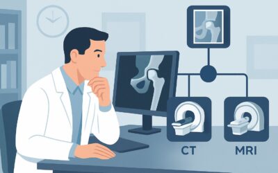
ACR-guided imaging workflow for Symptomatic hip arthroplasty patient, infection excluded. Additional imaging following radiographs. (Imaging after Total Hi

ACR-guided imaging workflow for Symptomatic hip arthroplasty patient, infection not excluded. Additional imaging following radiographs. (Imaging after Tota

ACR-guided imaging workflow for Symptomatic hip arthroplasty patient, history of acute injury. Additional imaging following radiographs. (Imaging after Tot
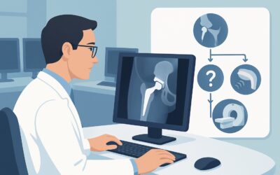
ACR-guided imaging workflow for Symptomatic patient with hip prosthesis. Initial imaging. (Imaging after Total Hip Arthroplasty): recommended study, differ

ACR-guided imaging workflow for Nonsuperficial (deep) soft tissue mass. Initial imaging. (Soft Tissue Masses): recommended study, differential, downstream

ACR-guided imaging workflow for Chronic extremity joint pain. Suspect erosive osteoarthritis. Radiographs normal or inconclusive. Next imaging study. (Chro
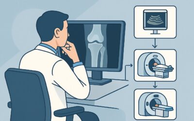
ACR-guided imaging workflow for Chronic extremity joint pain. Suspect calcium pyrophosphate dihydrate disease (pseudogout). Radiographs normal or inconclus

ACR-guided imaging workflow for Chronic extremity joint pain. Suspect gout. Radiographs normal or inconclusive. Next imaging study. (Chronic Extremity Join

ACR-guided imaging workflow for Chronic extremity joint pain. Suspect inflammatory arthritis (seropositive or seronegative arthritis). Radiographs normal o