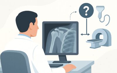
What Is the Next Imaging Step for an Obtunded Trauma Patient with a Negative Cervical Spine CT?

It’s 2 a.m. in the trauma bay. A patient, obtunded following a high-speed motor vehicle collision, has just returned from the scanner. The initial non-contrast Computed Tomography (CT) of the cervical spine is on your screen, and after a careful review, you see no evidence of fracture or malalignment. However, the patient cannot be examined clinically—they cannot report pain, and a reliable neurologic assessment is impossible. You cannot clear their cervical collar based on the CT alone. The critical question is what to do next to definitively rule out an unstable injury that could lead to catastrophic neurologic damage. This article details the American College of Radiology (ACR) Appropriateness Criteria workflow for this exact scenario, where the next recommended step, MRI cervical spine without IV contrast, is rated as May be appropriate (Disagreement), reflecting a complex and evolving standard of care.
Who Fits This Clinical Scenario for Post-Trauma Cervical Spine Imaging?
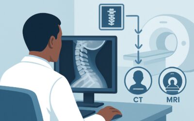
This guidance applies to a very specific and high-risk patient population. The inclusion criteria are precise: patients aged 16 years or older who have sustained acute blunt trauma and are obtunded, comatose, or otherwise unexaminable. A crucial prerequisite is that they have already undergone a technically adequate multidetector non-contrast CT of the cervical spine that was interpreted as negative for acute traumatic injury, such as a fracture or subluxation.
It is equally important to know who this workflow does not apply to, as similar presentations can lead to different imaging pathways:
- Alert and Examinable Patients: If a patient is awake, alert, not intoxicated, and has no distracting injuries, their cervical spine can often be cleared clinically using validated decision rules like the Canadian C-Spine Rule (CCR) or the National Emergency X-Radiography Utilization Study (NEXUS) low-risk criteria. These patients may not require any imaging at all.
- Patients with Known Fractures on CT: If the initial CT demonstrates a fracture, the workup shifts from screening for occult injury to characterizing the known injury and planning management. This often involves consultation with a spine specialist and may require additional imaging, but it follows a different diagnostic algorithm.
- Patients with High Suspicion for Vascular Injury: If the mechanism of injury or findings on the initial CT (e.g., a fracture extending through the transverse foramen) suggest a high risk for vertebral or carotid artery dissection, a dedicated vascular study like a CT Angiography (CTA) would be indicated. This represents a distinct clinical question focused on arterial injury rather than ligamentous stability.
What Diagnoses Are You Working Up After a Negative CT in an Obtunded Trauma Patient?
When a non-contrast CT of the cervical spine is negative for bony injury, the primary concern shifts to injuries of the soft tissues, which CT visualizes poorly. The subsequent imaging study is intended to identify clinically significant pathologies that could cause spinal instability or direct neurologic injury.
The most consequential diagnosis you are working up is a cervical ligamentous injury. The complex network of ligaments—including the anterior and posterior longitudinal ligaments, ligamentum flavum, and interspinous ligaments—is the primary stabilizer of the vertebral column. A complete disruption of this complex can render the spine unstable, even with intact bones. A missed unstable ligamentous injury could lead to spinal cord damage with subsequent movement. MRI is highly sensitive for detecting ligamentous edema and disruption.
Another critical consideration is Spinal Cord Injury Without Radiographic Abnormality (SCIWORA). In this condition, the patient suffers a direct injury to the spinal cord, such as a contusion, hemorrhage, or transection, without any evidence of fracture or malalignment on X-rays or CT. An obtunded patient cannot report symptoms like numbness or weakness, making direct imaging of the cord parenchyma with MRI essential for diagnosis and prognosis.
Less commonly, MRI may reveal an occult fracture not apparent on CT, particularly non-displaced fractures with significant surrounding bone marrow edema. It can also identify a traumatic epidural hematoma, which can cause cord compression and may be subtle or isodense to the cord on non-contrast CT.
Why Is MRI Cervical Spine Without Contrast Considered for a Negative CT?
The ACR rates MRI cervical spine without IV contrast as May be appropriate (Disagreement) for this scenario. This rating highlights an area of active debate in trauma care but underscores MRI’s unique diagnostic capabilities. The “Disagreement” reflects that while many centers consider MRI mandatory for clearing the C-spine in unexaminable patients, some protocols, citing the very low incidence of unstable injuries found after a negative modern multidetector CT, may clear the spine without it.
The rationale for proceeding with MRI is its superior soft-tissue contrast. It is the only imaging modality that can directly visualize the spinal ligaments, the spinal cord itself, the intervertebral discs, and the surrounding soft tissues. For the key differential diagnoses of ligamentous disruption and SCIWORA, MRI is the gold standard. The lack of ionizing radiation (0 mSv) is a significant advantage, especially in younger patients who may have already received a substantial dose from initial trauma imaging.
Ordering the study “without IV contrast” is a key specification. In the setting of acute trauma, gadolinium-based contrast agents are `Usually not appropriate` because they do not add significant diagnostic information for evaluating ligamentous integrity or primary cord contusion.
Alternative studies are rated lower for specific reasons:
- CTA head and neck with IV contrast is rated `Usually not appropriate` for this indication. While it is the right test for suspected vascular injury, it is not designed to evaluate the ligaments or spinal cord. It also delivers a significant radiation dose (☢☢☢ 1-10 mSv) and requires IV contrast.
- CT myelography cervical spine is also `Usually not appropriate`. This invasive test requires a lumbar puncture to inject contrast into the thecal sac and carries a very high radiation dose (☢☢☢☢ 10-30 mSv). It has been almost entirely replaced by non-invasive MRI for this clinical question.
Once you’ve decided on MRI cervical spine without IV contrast, our protocol guide covers the technique, contrast, and reading principles: MRI Cervical Spine Without Contrast.
What’s the Next Step After the Cervical Spine MRI Results?
The results of the cervical spine MRI will directly guide the next phase of management and determine whether the cervical collar can be removed.
- If the MRI is positive for an unstable ligamentous injury: This finding requires an immediate consultation with the neurosurgery or orthopedic spine service. The patient must remain in strict spinal immobilization. The definitive treatment is often surgical stabilization to prevent secondary spinal cord injury.
- If the MRI is positive for a spinal cord injury (e.g., contusion, edema, or hematoma): This also warrants urgent consultation with a spine specialist and neurologist. While management may be non-operative (e.g., blood pressure augmentation, supportive care), the finding is critical for prognostication and planning for long-term rehabilitation.
- If the MRI is negative: A technically adequate, high-quality negative MRI is widely considered sufficient to definitively rule out a clinically significant, unstable cervical spine injury. In this case, the cervical collar can be safely removed. This is a crucial step, as it helps prevent complications associated with prolonged immobilization, such as pressure ulcers, venous thromboembolism, and respiratory issues.
- If the MRI is indeterminate: In rare cases of an equivocal finding or a study degraded by motion artifact, the next step requires a multidisciplinary discussion between the clinical team, radiologists, and spine specialists. This may involve maintaining immobilization until the patient is clinically examinable or, in select cases, considering further imaging.
Common Pitfalls to Avoid in Clearing the Obtunded C-Spine
Navigating this clinical scenario requires careful attention to detail to avoid common errors.
Misinterpreting the “Disagreement” Rating: The ACR rating of “May be appropriate (Disagreement)” should not be interpreted as making the MRI optional or unnecessary. It signifies that institutional protocols vary, but it affirms that a negative CT alone is not sufficient to clear the spine in an unexaminable patient. The decision to proceed with MRI remains the most common and defended practice.
Prolonging Immobilization: While caution is paramount, unnecessarily delaying the MRI can lead to harm. If the patient is hemodynamically stable and has no other contraindications, the MRI should be performed in a timely manner to either identify injury or allow for safe collar removal.
Accepting a Suboptimal CT: The entire workflow is predicated on a high-quality, technically adequate initial CT. If the study is degraded by motion or streak artifact, it cannot be considered truly negative, and a repeat CT may be warranted before deciding on MRI.
If the MRI reveals an unstable fracture pattern, ligamentous disruption, or a compressive lesion like an epidural hematoma, the critical next step is to escalate immediately to the on-call spine service for emergent consultation.
Related ACR Topics and Tools
This article focuses on one specific clinical variant. For a comprehensive overview of imaging guidelines across all related scenarios, please consult our parent topic hub. You can also use the tools below to explore other ACR criteria, review imaging protocols, and discuss radiation dose with patients.
- For breadth across all scenarios in Acute Spinal Trauma, see our parent guide: Acute Spinal Trauma: ACR Appropriateness Decoded.
- ACR Appropriateness Criteria Lookup — for adjacent scenarios
- Imaging Protocol Library — for technique on the recommended study
- Radiation Dose Calculator — for cumulative dose conversations
Frequently Asked Questions
Why can’t I just clear the C-spine based on a negative high-quality multidetector CT?
Because CT is excellent for evaluating bones but is insensitive for detecting injuries to the spinal ligaments, spinal cord, or intervertebral discs. An obtunded patient cannot report pain or neurologic symptoms, and a purely ligamentous injury, which can cause catastrophic instability, will be missed on a CT scan.
What if my patient has a contraindication to MRI, such as an incompatible pacemaker?
This is a challenging situation that requires direct consultation with spine and radiology specialists. Alternatives are limited. Options may include keeping the patient immobilized until they are clinically examinable, or in very select cases, considering an invasive CT myelogram. The decision must be made on a case-by-case basis, weighing the risks of a missed injury against the risks of the alternative procedure.
The ACR rating is ‘May be appropriate (Disagreement)’. Does that mean some centers don’t perform the MRI?
Yes, this rating reflects an ongoing debate in trauma surgery. Some centers have protocols to clear the cervical spine based on a negative modern multidetector CT scan alone, citing the low incidence of unstable injuries found only on MRI. However, many institutions still consider MRI the standard of care to definitively rule out ligamentous injury before removing the collar in an unexaminable patient. The ‘Disagreement’ acknowledges this variation in practice.
Should I order the MRI with IV contrast?
No. For the specific question of acute ligamentous or spinal cord injury in trauma, the ACR rates MRI with IV contrast as ‘Usually not appropriate.’ Non-contrast sequences, including T1, T2, and fat-suppressed sequences like STIR, provide all the necessary information. Adding gadolinium-based contrast offers no significant benefit while increasing time, cost, and potential risk.
What if the obtunded patient also has a focal neurologic deficit but the CT is negative?
This patient still requires an urgent MRI of the cervical spine. However, the clinical context is different; you are now actively investigating a known neurologic deficit, not just screening for occult instability. The pre-test probability of a direct spinal cord injury (SCIWORA) is much higher, making the MRI even more critical for diagnosis and prognosis.
Reviewed by Pouyan Golshani, MD, Interventional Radiologist — May 29, 2026