

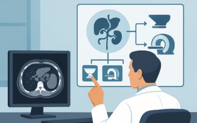
What Imaging Is Best for Jaundice with Suspected Mechanical Obstruction?
ACR-guided imaging workflow for Jaundice. Suspected mechanical obstruction based on initial imaging, clinical condition, or laboratory values. (Jaundice):
Part of the imaging decision system doctors were never taught to navigate at speed: which study to order, which protocol, and what the published appropriateness guidance actually says for GI imaging. Plain-English references, scenario by scenario.
ACR-guided imaging workflow for Jaundice. Suspected mechanical obstruction based on initial imaging, clinical condition, or laboratory values. (Jaundice):

ACR-guided imaging workflow for Jaundice. No known predisposing conditions. Initial imaging. (Jaundice): recommended study, differential, downstream pathwa
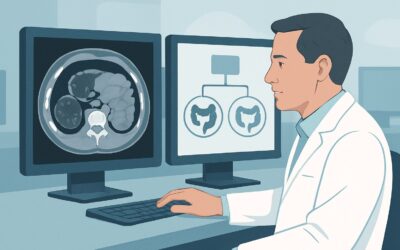
ACR-guided imaging workflow for Left lower quadrant pain. Suspected complication(s) of diverticulitis. Initial imaging. (Left Lower Quadrant Pain): recomme
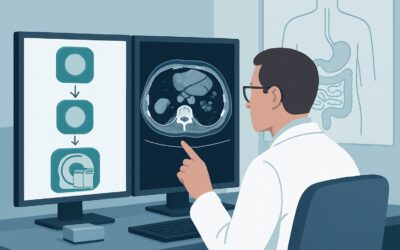
ACR-guided imaging workflow for Left lower quadrant pain. Suspected diverticulitis. Initial imaging. (Left Lower Quadrant Pain): recommended study, differe

ACR-guided imaging workflow for Delayed (greater than 1 month) postoperative development of dysphagia. Oropharyngeal or retrosternal. Initial imaging. (Dys

ACR-guided imaging workflow for Early postoperative dysphagia. Oropharyngeal or retrosternal. Initial imaging. (Dysphagia): recommended study, differential

ACR-guided imaging workflow for Right upper quadrant pain. Suspected acalculous cholecystitis. Negative or equivocal ultrasound. Next imaging study. (Right
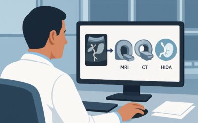
ACR-guided imaging workflow for Right upper quadrant pain. Fever, elevated WBC count. Suspected biliary disease. Negative or equivocal ultrasound. Next ima

ACR-guided imaging workflow for Right upper quadrant pain. No fever and no high white blood cell (WBC) count. Suspected biliary disease. Negative or equivo

ACR-guided imaging workflow for Right upper quadrant pain. Suspected biliary disease. Initial imaging. (Right Upper Quadrant Pain): recommended study, diff