


What Imaging Should You Order for Extremity Trauma in a Stable Polytrauma Patient?
ACR-guided imaging workflow for Adult. Major blunt trauma. Hemodynamically stable. Suspected extremity trauma. Initial imaging. (Major Blunt Trauma): recom
Part of the imaging decision system doctors were never taught to navigate at speed: which study to order, which protocol, and what the published appropriateness guidance actually says for MSK imaging. Plain-English references, scenario by scenario.
ACR-guided imaging workflow for Adult. Major blunt trauma. Hemodynamically stable. Suspected extremity trauma. Initial imaging. (Major Blunt Trauma): recom

ACR-guided imaging workflow for Adult. Major blunt trauma. Hemodynamically stable. Suspected facial injury. Initial imaging. (Major Blunt Trauma): recommen

ACR-guided imaging workflow for Adult. Major blunt trauma. Hemodynamically stable. Not otherwise specified. Initial imaging. (Major Blunt Trauma): recommen

ACR-guided imaging workflow for Adult. Major blunt trauma. Hemodynamically unstable. Initial imaging. (Major Blunt Trauma): recommended study, differential
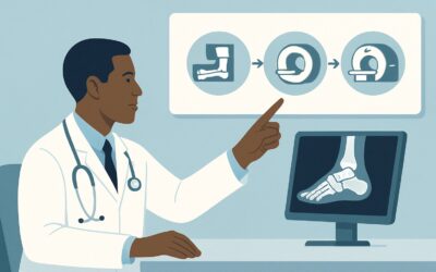
ACR-guided imaging workflow for Adult. Suspected osteomyelitis of the foot in patients with diabetes mellitus. Initial imaging. (Suspected Osteomyelitis of

ACR-guided imaging workflow for Suspect penetrating trauma with a foreign body in the soft tissues in the hand or wrist. Initial radiographs are negative.

ACR-guided imaging workflow for Initial radiographs showing metacarpophalangeal, proximal interphalangeal, or distal interphalangeal joint malalignment in

ACR-guided imaging workflow for Acute hand fracture on radiographs. Suspect hand tendon or ligament trauma. Next imaging study. (Acute Hand and Wrist Traum

ACR-guided imaging workflow for Initial radiographs showing distal radioulnar joint or carpal malalignment in the absence of fracture. Next imaging study.

ACR-guided imaging workflow for Acute wrist fracture on radiographs. Suspect wrist tendon or ligament trauma. Next imaging study. (Acute Hand and Wrist Tra